
X-Ray
Dental X-rays are an essential part of maintaining good oral health, providing a clear and detailed view of areas that cannot be seen during a regular dental exam, and aiding in the early detection and treatment of dental problems.
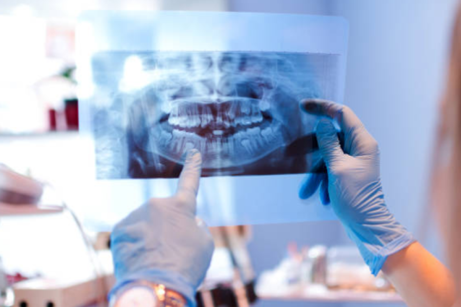
Dental X-rays, also known as radiographs, are diagnostic tools used to capture images of the teeth, bones, and soft tissues inside the mouth.
They help dentists identify problems that are not visible during a regular dental examination, such as cavities, tooth decay, impacted teeth, bone loss, abscesses, cysts, and other dental issues.
Dental X-rays are a crucial part of preventive care and are essential for accurate diagnosis and treatment planning.
Process:
- Preparation: Before the X-ray, the patient will be asked to remove any jewelry, eyeglasses, or removable dental appliances that could interfere with the imaging process. A lead apron or thyroid collar is placed over the patient’s body to protect against any exposure to radiation.
- Positioning: The dental technician or assistant will position the patient comfortably in the dental chair. The X-ray machine is adjusted to target the specific area of the mouth that requires imaging. The patient may be asked to bite down on a small piece of plastic or hold a sensor in place to capture the image.
- Taking the X-ray:
- The X-ray machine is activated, and a small, controlled amount of radiation is used to capture images of the teeth and surrounding structures.
- Depending on the type of X-ray being taken, the patient may need to reposition or adjust their head slightly. Types of dental X-rays include bitewing, periapical, panoramic, and occlusal, each designed to capture different views of the mouth.
- The process is quick, painless, and usually takes just a few seconds for each image.
- Image Processing: The captured X-ray images are processed either digitally or on film. Digital X-rays provide almost instant results and use less radiation compared to traditional film X-rays. The images are displayed on a computer screen, allowing the dentist to view them immediately.
- Diagnosis and Treatment Planning: The dentist reviews the X-ray images to assess the patient’s dental health, identify any hidden issues, and develop a comprehensive treatment plan. X-rays are vital for detecting problems early, which allows for more effective and less invasive treatment options.
- Follow-Up: The dentist will discuss the findings with the patient, explaining any concerns and recommending necessary treatments or procedures. The frequency of dental X-rays will depend on the patient’s age, dental history, and specific dental health needs.